
OCT scan: look deeper into your eye
A photograph shows the surface. An OCT scan reveals the layers beneath it. Discover how our Topcon Triton helps assess your retina and optic nerve at Sammy Optiek in Zuilen, Utrecht.
Swept-Source · 3D OCT
Safe, contact-free and usually without drops
Structural OCT uses light rather than X-rays. It does not touch the eye and needs no injection. There are no known harmful side effects from the scan itself when the equipment is used normally.
Our Topcon Triton can image the retina without dilating drops. They are generally unnecessary for a routine OCT acquisition, a convenient benefit. Small pupils, a less clear lens or additional eye examinations may still lead the optometrist to recommend drops. We discuss this beforehand.
What is an OCT scan?
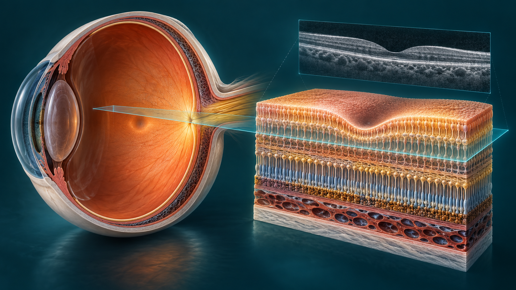
OCT stands for optical coherence tomography. Reflected light creates thin cross-sections of tissue, without X-rays or touching your eye. The optometrist can examine retinal structure and assess changes in thickness or shape.
A sight test mainly checks how clearly you see and which prescription you need. A retinal photograph shows the back of the eye from above. OCT adds depth: layers, fluid and distortion may become visible. Symptoms and other eye examinations give a finding its meaning.
A thin layer with a remarkable job
The retina is the light-sensitive lining at the back of your eye. It converts light into signals for your brain. Layers and distances are simplified in our enlarged illustrations.
Macula and fovea
The macula supports reading, recognising faces and fine detail. At its centre is the fovea, the small depression visible in many OCT sections.
Photoreceptors and pigment layer
Rods and cones detect light. Behind them lies the retinal pigment epithelium (RPE), which supports these cells. Changes around these layers may affect central vision.
Nerve fibres and optic nerve
Nerve cells send signals to the brain through the optic nerve. OCT helps assess the nerve fibre layer; a thin layer alone does not prove glaucoma.
Vitreous and choroid
The vitreous lies in front of the retina and may pull on it. The choroid lies behind it and supplies the outer retinal layers with oxygen and nutrients.
Our Topcon Triton: structural depth in 3D
Sammy Optiek uses the Topcon DRI OCT Triton, a Swept-Source OCT. Many light measurements build a volume. Depending on the scan protocol and image quality, it can help assess retinal layers, the vitreoretinal interface, the macula and the area around the optic nerve. Deeper structures such as the choroid may also be visible.
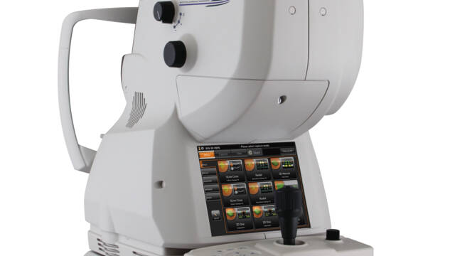
Our Triton 3D OCT in action
Explore the examination and retinal images in 30 seconds.
From a cross-section to a volume
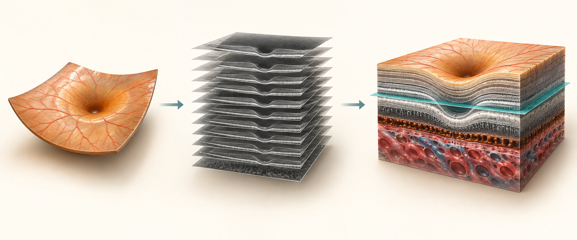
An A-scan measures depth. A row of these measurements forms a B-scan, or cross-section. Combining adjacent sections creates a 3D volume. The optometrist can scroll through an area, locate changes and compare thickness measurements where appropriate. A 3D view reconstructs measurements; it is not a photograph of individual cells.
Explore the retina in real OCT images
Choose an example, compare it with a healthy reference and zoom into the real scan.
Drusen / dry AMD
The bumps below the outer retinal layers are drusen. The markings are part of the published scan.
Published examples from different eyes and devices; magnification and scale vary. One scan does not establish a diagnosis. Cropping and zoom preserve the original pixels.
Explore the explanatory 3D model
Retina explorer
Choose an example, move through the sections and turn the volume. This simplified model explains patterns; it does not assess your eyes.
Healthy reference
A gradual foveal depression and continuous layers. Normal shape and thickness vary between eyes. This diagram does not establish eye health.
Interactive diagram · not a real OCT · not to scale
Why primary eye care screening matters
Reading an eye chart clearly does not necessarily mean every eye structure is healthy. Some changes cause few symptoms at first. An accessible optometrist can investigate whether a complaint relates to prescription or needs further eye-health assessment. OCT may add useful information.
At Sammy Optiek, Marc, our optometrist, interprets the scan alongside your history and eye examination. Primary screening helps decide the next step: explanation, review or referral to an ophthalmologist. A scan does not treat disease or guarantee its prevention.
Not everyone needs OCT at fixed intervals. Its value depends on symptoms, earlier findings and individual risk. A normal OCT does not exclude every eye condition: a central scan does not examine the entire peripheral retina.
When may OCT be useful?
- Unexplained blurred or distorted central vision. Seek medical advice first for new or sudden symptoms.
- Macular assessment when changes such as fluid or macular degeneration are suspected.
- Additional nerve-fibre assessment for suspected glaucoma, alongside other examinations.
- A baseline or comparison of known changes when advised by your clinician.
Results need professional interpretation
A colour on a thickness map is not a diagnosis. Age, high myopia, scan position, movement and errors in automated layer boundaries can affect measurements. Uncertain images may be reviewed or repeated. OCT does not replace your agreed diabetic retinal screening or existing ophthalmologist appointments.
OCT image atlas: what can you see?
Real educational images from public sources, acquired on different devices. These are not scans of Sammy clients and do not predict your results. Select an image to enlarge it. Each has its source and licence; originals are unchanged.
Macular hole beside a reference
Compare the continuous central contour on the left with the opening in the macula on the right.
Source: Betty Wills (Atsme) · CC BY-SA 4.0
Original, unchanged
Small macular hole
Even a small central break may matter. The optometrist examines several sections.
Source: Hanay · CC BY-SA 3.0
Original, unchanged
Adhesion, traction and macular hole
Examples distinguish vitreous attachment, pulling with distortion and a hole.
Source: Yoanmb · CC BY-SA 4.0
Original, unchanged
Cystoid macular oedema
Dark spaces within the thickened retina illustrate cystoid fluid accumulation.
Source: Rebellos · CC BY-SA 3.0
Original, unchanged
Diabetic macular oedema
This teaching example shows fluid and altered macular structure in diabetes. OCT does not replace diabetic screening.
Source: Jmarchn · CC BY-SA 3.0
Original, unchanged
Oedema in cross-section and 3D
A volume shows the extent of an elevated area; the section reveals internal structure.
Source: Jmarchn · CC BY-SA 3.0
Original, unchanged
A 3D thickness view
Height and colours show thickness differences. Scale, scan protocol and layer boundaries are needed for interpretation.
Source: Jmarchn · CC BY-SA 3.0
Original, unchanged
Drusen in macular degeneration
Annotations in this source image show drusen: elevations beneath the pigment layer. OCT shows structure, not a blood-flow map.
Source: Yoanmb · CC BY-SA 4.0
Original, unchanged
Epiretinal membrane
A membrane on the inner surface can distort the foveal contour. Vision and symptoms also matter.
Source: Jmarchn · CC BY-SA 3.0
Original, unchanged
What happens during your OCT examination?
Your question and history
Tell us what has changed, when it started, your medication and previous eye examinations. Bring your glasses and any earlier reports.
Look steadily at a target
Your chin and forehead rest on supports while you look at a fixation point. Acquisition is usually brief; discussion and assessment take additional time.
Images in context
The optometrist checks quality and interprets the scan with other findings. A routine OCT acquisition usually needs no drops. Extra images or dilating drops may be recommended for additional examinations.
A clear next step
We explain findings and advice. If disease is suspected, we discuss appropriate referral and urgency.
3D OCT at Sammy Optiek
A 3D OCT scan at Sammy Optiek costs €79. Use the online appointment tool to choose Optometry; your scan request is carried through. The optometrist decides which examination suits your question. Ask about additional examination costs beforehand and check reimbursement with your insurer.
Doornburglaan 13 · Zuilen · Utrecht
Book an Optometry appointmentFrequently asked questions
Does an OCT scan hurt?
It does not touch the eye and is generally painless. You need to sit still and look at a target briefly.
Does the Triton scan need eye drops?
A routine OCT acquisition generally needs no dilating drops. Sometimes the optometrist recommends them for image quality or additional examination. Dilation may temporarily blur vision and cause light sensitivity. Arrange transport if needed and do not drive while vision is inadequate.
Is a normal OCT a guarantee?
No. OCT examines specific structures in the scanned area. A normal scan does not exclude every condition or replace the rest of the eye examination.
Can OCT diagnose glaucoma?
It can show relevant nerve-fibre changes. Diagnosis also considers pressure, optic nerve and visual fields.
How often do I need a scan?
It depends on risks, symptoms and findings. Your optometrist or ophthalmologist recommends follow-up; there is no universal schedule.
Read more from reliable sources
Original patient information prepared for Sammy Optiek. Sources consulted on 7 October 2026. This is general education; discuss your own results with your optometrist.
- Oogartsen.nl · OCT examination ↗
- Cleveland Clinic · OCT: examination and results ↗
- Topcon Healthcare · Triton specifications and options ↗
- Optometristen Vereniging Nederland · Optometry in primary care ↗
- National Eye Institute · Macular degeneration ↗
- National Eye Institute · Diabetic retinopathy ↗
- National Eye Institute · Glaucoma and screening ↗
- Moorfields Eye Hospital · Epiretinal membrane ↗
- Moorfields Eye Hospital · Central serous retinopathy ↗
- Thuisarts · Flashes and urgent symptoms ↗